The Skeletal scintigraphy is a modern imaging procedure that is particularly useful in the Equine orthopaedics has proven itself.
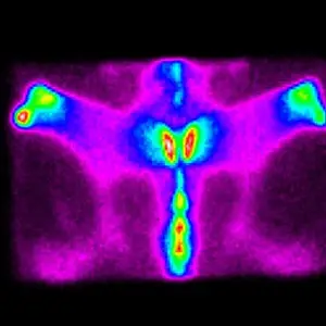
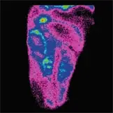
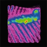
It enables the display of Foci of inflammation and Changes in bone metabolism - often before they become visible in the X-ray image.
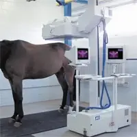
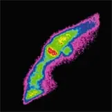
An approved radioactive tracer is administered intravenously. Areas of inflammation or increased osteoblastic activity demonstrate increased tracer uptake, which can be accurately visualised using a highly sensitive gamma camera.
The Pferdeklinik Bargteheide has many years of experience in this area:
Already since 1996 we use skeletal scintigraphy on a standing horse in.
Today we can up to 15 horses per week with this method.
Indications for Skeletal Scintigraphy
- Performance and rideability problems with suspected pathology of the cervical spine or thoracolumbar region
- Suspected fissures, stress fractures or fractures in anatomically challenging regions (e.g. pelvis, hip, femur)
- Unexplained or severe lameness
- Enthesopathies, such as lesions at the origin of the suspensory ligament
- Monitoring and follow-up of previously diagnosed orthopaedic conditions
- Assessment of suspected perfusion or vascular disorders
- Joint pathology not detectable on conventional radiographic examination
- Evaluation of uncooperative patients as an alternative to diagnostic nerve or joint blocks
Your Benefits at a Glance
- Examination of the entire skeleton or selected anatomical regions
- Detection of lesions in areas that are difficult to access or radiographically inconspicuous
- Early identification of pathological changes before structural damage becomes apparent
- Performed in the standing, sedated horse – no general anaesthesia required
Following skeletal scintigraphy, more targeted diagnostic procedures such as radiography, ultrasonography, diagnostic anaesthesia, MRI or CT can often be performed to further characterise the findings and guide treatment planning.