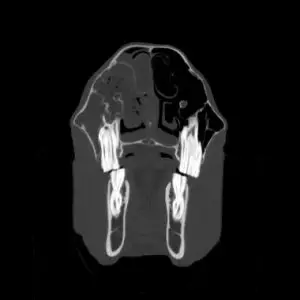
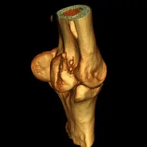
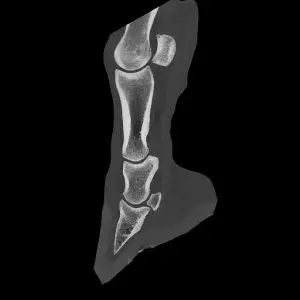
Computed tomography, like magnetic resonance imaging, is a cross-sectional imaging procedure. Unlike MRI, CT images are produced with the help of X-rays. The horse is scanned layer by layer, a computer then assembles these countless individual images into a detailed three-dimensional image of the examined region. This allows the detection of minor osseous and soft-tissue abnormalities – findings that would otherwise remain undetected on conventional radiographic images. If necessary, a contrast agent can be used to visualise certain structures even better.
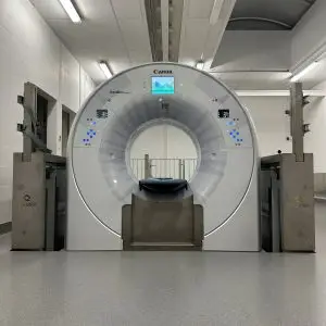
Our powerful CT scanner (Qualibra Exeed, equipped with Aquilion Exceed from Canon Medical and artificial intelligence from AiCE) with a particularly large opening (gantry) of 90cm enables the visualisation of previously inaccessible body regions such as the pelvis, shoulder region and other parts of the spine.
A key advantage of our system is the ability to perform many examinations on a standing, sedated horse. Thanks to a specially designed pit that allows the CT unit to move not only forwards and backwards but also vertically, we are able to examine the head, cranial sections of the cervical spine and the distal limbs (up to the carpal/ tarsal region) without the need for general anaesthesia. This reduces the risk for the horse and enables the procedure to be carried out with significantly less effort.
Computed tomography examinations of the upper limb and the more caudal sections of the cervical spine do require general anaesthesia. In these cases, the horses are anaesthetised as they would be for surgery and can the be transported directly through the operation room into the adjacent CT room. This setup ensures a smooth anaesthetic process and also enables surgical procedure to be performed under CT guidance if necessary.