In ultrasound imaging, sound waves are almost completely reflected by bone and air, which means that sonography is mainly used to visualize soft tissue structures such as tendons, ligaments, blood vessels, and internal organs. However, the surface of bones can also be examined with high resolution. X-rays and ultrasound are therefore two complementary procedures. The specialized techniques of Doppler sonography and color Doppler sonography provide additional information about the direction, speed, and strength of blood flow in the heart and blood vessels. These procedures are primarily used in cardiac examinations.
Die besonderen Verfahren der Doppler-Sonografie und der Farbdoppler-Sonografie liefern zusätzlich Informationen über die Strömungsrichtung, -geschwindigkeit und -stärke eines Blutflusses im Herzen und in den Gefäßen. Diese Verfahren finden vor allem bei der Herzuntersuchung Verwendung.
Both a modern ultrasound system and an experienced veterinarian with detailed knowledge of the anatomical region are essential for a highquality ultrasound examination. The preparation of the patient is also critical: the horse must be able to stand quietly to allow precise imaging and reliable diagnostic assessment. Because the clinic represents an unfamiliar and potentially stressful environment, many horses are more nervous than in their home stables, so sedation is often required for the examination. The region to be assessed is clipped, cleansed, and coated with ultrasound gel to optimize the coupling between the transducer and the skin. Multiple highperformance ultrasound systems with different transducers are available to accommodate the various clinical indications and examination regions.
Areas of application of sonography in horses:
The areas of application of sonography are diverse and have developed considerably in recent years. The constantly improving technology and increasing expertise enable examinations in many areas of equine medicine.
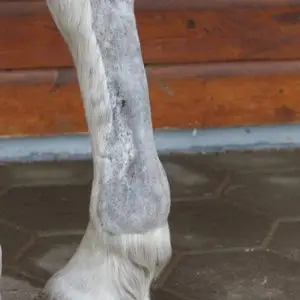
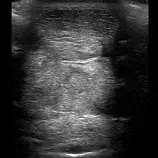
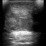
Sonography of the tendons and ligaments:
Ultrasound examination of the flexor tendons is one of the most important procedures in equine orthopaedics. Highresolution transducers allow even very small defects in the tendons to be detected. Precise localisation and assessment of the lesion not only enables the development of an individual therapy plan, but also targeted treatment. For example, regenerationpromoting medication can be injected directly into the defect under ultrasound guidance. The healing process can be monitored by followup examinations every six to eight weeks, and the exercise programme can be adjusted accordingly.
Sonography of joints:
Under ultrasound guidance, injection needles can be placed precisely into diseased or difficult-to-access tissues, such as tendon lesions or facet joints in the cervical spine. This technique also enables targeted needle biopsies, for example from the live
Ultrasound-guided injections:
Under ultrasound guidance, injection needles can be placed precisely into diseased or difficult-to-access tissues, such as tendon lesions or facet joints in the cervical spine. This technique also enables targeted needle biopsies, for example from the live.
Sonography for acute injuries:
Ultrasound can be used to identify which structures (joints or tendon sheaths) are affected. Foreign bodies such as wood splinters, which are not visible on radiographs, can also be detected using ultrasound.
Use in foal medicine:
Ultrasound is now indispensable in foal medicine. As a rectal examination is not possible in cases of colic, ultrasound provides a window into the abdominal cavity and can indicate possible causes.
Visualisation of internal organs such as the internal umbilical structures, intestines, kidneys and bladder, as well as free fluid in the abdomen (for example in the case of bladder rupture), supports diagnosis. Ultrasound also enables early detection of pneumonia and monitoring of the healing process.
Gynaecological ultrasound:
In gynaecological ultrasound, the probe is inserted rectally, allowing visualisation of the ovaries and uterus. This examination is primarily used to determine cycle status, in particular to identify the optimum time for insemination and to detect early pregnancy. Changes or abnormalities in the ovaries and uterus can also be visualised in this way.
Ultrasound of the lungs:
Healthy lungs are filled with air, so on ultrasound usually only the smooth pleural surface is visible as a bright linear echo. In cases of pneumonia, however, certain areas of the lung are no longer aerated and can be easily visualised. Accumulations of fluid in the chest cavity (pleural effusion) can also be identified using sonography.
Echocardiography:
During echocardiography, the cardiac chambers, myocardium and heart valves are examined. Both the size of the chambers and the motion patterns of the heart muscle can be assessed precisely. Using Doppler ultrasound, blood flow across the individual valves can be visualised and valvular defects can therefore be evaluated (see heart examination).
Sonography of the abdomen:
Ultrasound examination of the abdomen includes assessment of the liver, spleen, kidneys, urinary bladder and parts of the intestine. In some cases, it can also provide valuable additional information in the investigation of colic.
Sonography of the eye:
If the lens or cornea are opaque, the interior of the eye can no longer be inspected directly. In these cases, the posterior segments of the eye can still be evaluated using ultrasound.
Examination of neck and back
Suspected disease in the neck region usually involves changes in the soft tissues, which is why ultrasound is the preferred diagnostic modality. Assessment of the facet joints in the cervical spine is only possible to a limited extent with ultrasound; in such cases, radiography in combination with scintigraphy is preferred.
Treatment of the facet joints by local injection can only be carried out under ultrasound guidance because of the very narrow joint spaces. The muscles, ligaments and small vertebral joints of the lumbar spine can be visualised in the back region. However, the sacroiliac joint can only be imaged in a very limited area.
Examination of the pelvis:
Examination of the pelvis in horses is often challenging and in some cases only possible under general anaesthesia. Ultrasound is less invasive and allows visualisation of parts of the hip joint, the bony surface of the pelvis and, for example, pelvic fractures, with a penetration depth of up to 30 cm. The caudal portion of the sacroiliac joint can be examined using transrectal ultrasound.
Examination of vessels:
Ultrasound enables visualisation of thrombosis in the jugular veins or limb arteries. It is used to diagnose vascular stenoses or occlusions.